|
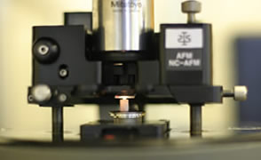
Atomic Force Microscopy
(AFM) is a form of scanning probe microscopy. The AFM probe has a very sharp tip, at the end of a small cantilever beam. The probe is attached to a piezoelectric scanner tube, which scans the probe across a selected area of the sample surface. Interatomic forces between the probe tip and the sample surface cause the cantilever to deflect as the sample's surface topography (or other properties) changes. A laser light reflected from the back of the cantilever measures the deflection of the cantilever, and this information is fed back to a computer, which generates a map of topography and/or other properties of interest. Areas as large as about 100 microns square to less than 100 nm square can be imaged.
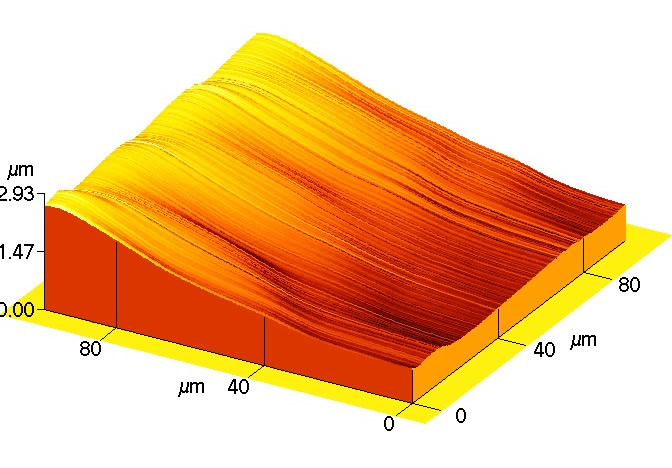
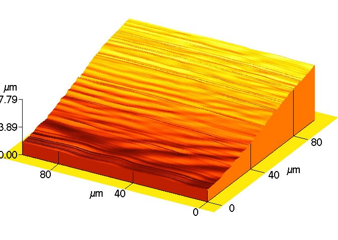
Atomic force microscopy was used to compare the sealing surfaces of two components manufactured fromdiffernt cavities within a multi cavity tool. One cavity produced poor seal quality whilst the other produced a good, consistent seal.
AFM images of the two surfaces are shown below:
 |
 |
| Figure 1. AFM image from cavity showing good seal quality. |
Figure 2. AFM image from cavity showing poor seal quality |
As can be seen from these images, whilst the component from the good cavity shows a relatively smooth surface, the cavity producing the poor seal quality has a rippled, undulating surface. This cavity, with a reduced tool temeprtaure produces stick/slip flow in the critical region of the sealing surface.
|